Throughout the gastrointestinal tract, subserve 2 primary function
- Digestive enzymes are mostly secreted in the entire alimentary canal, from the mouth to the anus.
- Mucosal glands from the mouth to the anus provide mucus lubrication and protection of all parts of the alimentary tract.
Majorly digestive secretions are made in response to the food which is sitting in the alimentary canal, and the quantity secreted in each segment of the track is almost exactly the amount needed for proper digestion.
Furthermore, in some portions of the gastrointestinal tract, even the type of enzymes and other constituents of the secretions are varied by the types of food present.
General Principle of Alimentary Tract Secretions
- MUCOUS CELLS/GOBLET CELLS—–function in response to local irritation of epithelium; they extrude mucus directly onto the epithelial surface to act as a lubricant that also protects the surface from excoriation and digestion.
- (pits that represent invaginations)CRYPTS OF LIEBERKUHN IN SMALL INTESTINE —–are deep and contain specialised secretory cells.
- OXYNTIC GLAND/TUBULAR GLAND IN STOMACH AND UPPER DUODENUM—–shows an acid- and pepsinogen-secreting gland of the stomach.
- SALIVARY GLAND
- PANCREAS
- LIVER
BASIC MECHANISM OF STIMULATION OF THE ALIMENTARY TRACT GLANDS

effect of contact of food with the epithelium function of the enteric nervous stimuli.
- The mechanical presence of food in the gastrointestinal tract usually causes the glands of that region and often of the adjacent regions to secrete moderate to large quantities of juices.
- Part of this local effect, especially the secretion of mucus by mucous cells, results from direct contact stimulation of the surface glandular cell by the food.
- In addition, local epithelial stimulation also activates the enteric nervous system of the gut wall.
- Types of stimuli that do this are
- TACTILE STIMULATION
- CHEMICAL IRRITATION
- DISTENTION OF THE GUT WALL
- The resulting nervous reflexes start to stimulate mucous cells on the gut epithelial surface and the deep glands in the gut wall henceforth increasing their secretion.
AUTONOMIC STIMULATION OF SECRETION
PARASYMPATHETIC STIMULATION
- Stimulation of parasympathetic nerves to the alimentary tract almost invariably increases the rate of alimentary glandular secretion
- Majory is true for the glands in the upper portion of the tract innervated by the glossopharyngeal and vagus parasympathetic nerves
- These glands are the salivary gland, oesophageal gland, gastric glands, pancreas, and Brunner’s gland in the duodenum.
SYMPATHETIC STIMULATION
- sympathetic nerves which are going to the gastrointestinal tract when stimulated cause a little to moderate increase in secretion by some of the local glands.
- But sympathetic stimulation also results in the constriction of the blood vessel that supplies the glands.
- therefore, sympathetic stimulation has a dual effect
- sympathetic stimulation alone usually slightly increases the secretion
- if parasympathetic or hormonal stimulation is already causing an enormous amount of secretion by the glands, superimposed sympathetic stimulation usually lowers the secretion sometimes significantly, mainly because of vasoconstrictive reduction of the blood supply.
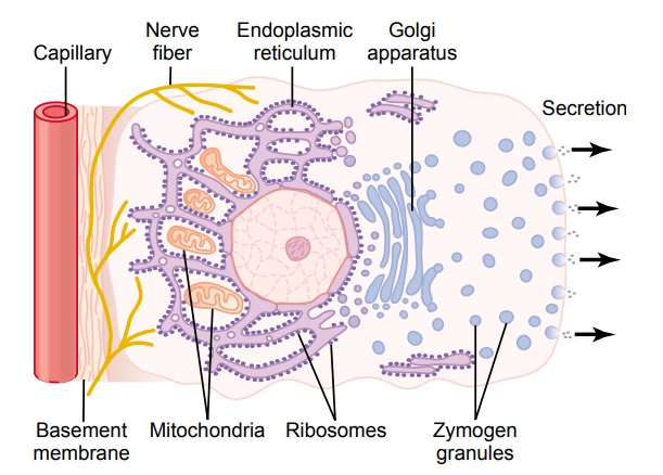
BASIC MECHANISM OF SECRETION BY GLANDULAR CELLS

Secretion of organic substances:
- Nutrient material needed for the formation of the secretion must first diffuse or be actively transported by the blood in capillaries into the base of the glandular cell.
- a number of mitochondria that are located inside the glandular cell near its base use oxidative energy to form ATP i.e adenosine triphosphate.
- The energy provided by ATP, n addition to appropriate substrates provided by the nutrients, is then used to synthesise the organic secretory substances;
This synthesis occurs completely in the Endoplasmic Reticulum and Golgi Complex of the glandular cell. Ribosomes(RER—-Rough endoplasmic reticulum which has ribosomes attached) adherent to the reticulum is specifically responsible for the formation of the protein that is secreted.
- The Secretory materials are transported through the tubules of the endoplasmic reticulum, passing in about 20 min to the vesicles of the Golgi complex.
- materials are modified, concentrated, added to, and then discharged into the cytoplasm in the form of the secretory vesicle, which is stored in the apical end of secretory cells.
- These secretory vesicles are still stored until nervous or hormonal control signals cause the cells to extrude the vesicular contents through the cell surface
~this is done by following ways
* the control signal first incrrease the
cell membrane permiability to Ca+
ions and calcium eners the cell
* Ca+ causes many of the vesicle to
fusewith the apical cell membrane.
*Then the apical cell membrane breaks
open,thus emptying the vesicle to the
exterior;called as EXOCYTOSIS.
WATER AND ELECTROLYTE SECRETION

- The second necessary for glandular secretion is the secretion of sufficient water and electrolyte to go along with the organic substances.
- Nerve stimulation has a specific effect on the basal portion of the cell membrane to cause the active transport of chloride ions to the cell interior.
- the resulting increase in electronegativity induced inside the cell by excessive negatively charged chloride ions (Cl-), then cause positive ion such as sodium(Na+) also to move through the cell membrane to the interior of the cell.
- The excess of both + and – ions inside the cell creates an osmotic force that causes osmotic pressure inside the cell. therefore increasing cell volume and hydrostatic pressure inside the cell, causing the cell itself to swell up.
- The pressure in the cell results in the initiation of a minute opening of the secretory border of the cell causing flushing of water, electrolyte and organic materials out of the secretory end of the glandular cell.
- (the nerve ending on the glandular cells are principally on the bass of the cells
- Microelectrode studies show that the normal electrical potential across the membrane at the base of a cell is between is 30 millivolts to 40 millivolts negatively on the interior and positively on exterior
- Parasympathetic nerve stimulation increases this polarisation voltage value by 10 – 20 millivolts more negative than usual)
LUBRICATING AND PROTECTIVE PROPERTIES OF MUCUS, AND THE IMPORTANCE OF MUCUS IN THE GASTROINTESTINAL TRACT
- Mucus is a thick secretion composed mainly of water, electrolytes and a mixture of several glycoproteins, which themselves are composed of large polysaccharides bound with much smaller quantities of protein
- Mucus is slightly different in parts of the gastrointestinal tract, but everywhere it has several important characteristics that make it both an excellent lubricant and a protectant wall.
- Mucus has adherent qualities that make it adhere tightly to the food or other particles and spread as a thin film over the surfaces.
- It has a sufficient body that coats the wall of the gut and prevents actual contact of most food particles with the mucosa.
- Mucous has low resistance for slippage so that the particles can slide along the epithelium with great ease.
- The mucus causes the faecal particle to adhere to one another to form faeces that are expelled during a bowel movement.
- Mucus is immensely resistant to digestion by the gastrointestinal enzymes.
- The glycoproteins of mucus have amphoteric nature which means that they are capable of buffering small amounts of acids/(base) alkalies; also mucus might contain a considerable amount of bicarbonate ions which specifically neutralize acids.